



| 货号 | 规格 | 价格 | 上海 | 北京 | 珠海 | 重庆 | 武汉 | 购买数量 |
|---|---|---|---|---|---|---|---|---|
R43295-5reactions促销 |
BioReagent | ¥1300.00 ¥1170.00 |
10 | - | - | - | - |
|
荧光染料抗体标记试剂盒设计注重效率与可靠性,可在60分钟内快速完成标记,仅需极少的人工操作时间。试剂盒内含抗体标记所需的所有试剂,包括NHS酯活化染料、缓冲液和脱盐柱。试剂盒中的活性染料可与抗体或蛋白质中的赖氨酸残基高效结合,生成共价连接的荧光传感器。配套的脱盐柱可去除过量未反应的染料,回收率可达70%-95%。试剂盒提供5管活性染料,每管活性染料都含有优化量的染料,可实现100μg抗体的标记反应。我们提供多种颜色染料试剂盒,包可兼容多种检测系统,包括荧光显微镜和流式细胞术。产品组分A1491934Component 5 reactionsStorageQuantity Per reactionA1491934AAF488 NHS Ester5 vials-20℃. Store in the dark. 1 vial for labeling 100 μg of antibodyA1491934BNaHCO3100 mgRT.Prepare according to instructionsA1491934CSpin Desalting Column5 EA2-8℃. Do not freeze.1 EA for 1 reactionA1491934DCollection Tubes10 EART.2 EA for 1 reaction其他所需试剂1. 离心机2. 用于标记的所需抗体(不含BSA或任何载体蛋白)。3. PBS缓冲液(pH 7.2-7.4)。重要须知1. 纯化的抗体缓冲液中不得含有铵离子或者伯胺组分,例如Tris、甘氨酸、乙醇胺、三乙胺、谷胱甘肽或咪唑,所有这些物质都会显著抑制蛋白质标记。2. 用于标记的抗体需经过纯化,不含有BSA或明胶等稳定剂。3. 抗体浓度1-2mg/mL最佳,浓度低于1mg/mL, 标记效率可能会降低,浓度过高,抗体体积会过少,不利于纯化回收。4. 脱盐柱建议一次性使用。5. 试剂盒使用之前,需将所有组分恢复至室温。6. 荧光染料均存在淬灭问题,请尽量注意避光,以减缓荧光的淬灭。使用说明1. 缓冲液配制:a) 1M NaHCO₃的配制。根据样品量配制适量的1M NaHCO₃溶液,如称取42mg NaHCO₃,加入0.5mL超纯水即得1M NaHCO₃溶液。NaHCO₃可维持标记反应体系的pH在7-9之间,提高标记效率。注:NaHCO₃溶液须现用现配。2. 抗体标记a) 如果待标记的抗体浓度≥1mg/mL,并且在适当的缓冲液中,则将其稀释至1mg/mL,并加入10%抗体体积的1M NaHCO₃缓冲液。如果抗体是从适当的缓冲液中冻干的粉末,通过向抗体中加入适量的0.1M NaHCO₃缓冲液来制备1mg/mL的抗体溶液。用去离子水将1M溶液稀释10倍,制备0.1M NaHCO₃缓冲液。b) 将100μL抗体溶液转移到活性染料管中。盖上管盖,轻轻翻转几次,使染料完全溶解。注意:为确保染料完全溶解,请将标签从活性染料管上撕下。c) 将反应管室温避光反应1h。反应可以在振荡器或混合器上进行,建议上下翻转速度为25rpm。如果在反应过程中不使用混合仪器,则应每10分钟将反应溶液倒置混合一次。注意:反应进行期间,可提前进行步骤3的操作,完成脱盐柱的平衡。3. 脱盐柱预平衡a) 松开脱盐柱盖子,折断柱子下端堵头,将脱盐柱置于配套的2mL收集管中。1000xg离心2分钟,丢弃离心下来的保存溶液,并将脱盐柱放回收集管中。注意:使用非水平转头的情况下,由于离心会使树脂压实形成一个向上的斜面,该斜面的方向宜在后续步骤中保持,所以在脱盐柱外壳上的斜面向上位置做标记,在随后的离心步骤中需要调整好离心管的放入方向,确保离心后斜面的方向和位置不会改变。b) 向脱盐柱树脂顶部加入500μL PBS,轻轻拧上脱盐柱上盖,1000×g离心2分钟,丢弃溶液,重复此步骤3次。4. 荧光标记抗体纯化a) 将平衡后的脱盐柱转移至新的收集管中。小心的将反应管中的反应液加入脱盐柱中,盖上脱盐柱盖子,注意盖子轻轻拧上即可。b) 脱盐柱1000xg离心2分钟,收集管中的流出即为所需收集的抗体偶联物。5. 测定蛋白质浓度和DOL(可选)DOL(标记度)是衡量荧光标记效率的关键参数,表示荧光团与目标分子(如抗体或蛋白质)的摩尔比。可以从抗体偶联物的吸收光谱中测定。a) 检测标记产物280nm的吸光度(A₂₈₀)和对应染料最大吸收波长处的吸光度(Adye)。不同染料的参数可参考下面表格。Cat.No.Dyesλmax (nm)Em (nm)εdye (M⁻¹cm⁻¹)CF₂₈₀Target DOLMW (g/mol)A1491932AF35034344119,0000.194-9410A1491933AF40540142234,0000.74-91028A1491934AF48849052576,0000.14-91081A1491935AF53253055581,0000.094-9723A1491936AF555553568150,0000.084-81250A1491937AF59459061892,0000.564-9820A1491938AF647650668239,0000.034-81300A1491939AF660663691132,0000.13-81100A1491940AF680681704184,0000.053-71150A1491941AF700696719192,0000.073-71400A1491942AF750752776240,0000.043-81300F1491944FITC 49851773,0000.353-8389P1491945Pacific Blue41045546,0000.24-9339O1491947Oregon Green 48849852676,0000.123-8509T1491948TRITC557576100,0000.341-3443C1491949Cy3554566150,0000.073-5734C1491950Cy5647665250,0000.033-5754b) 计算抗体偶联物中抗体浓度:抗体浓度(mg/mL) = (A280 - CF280 × Adye) / 1.4c) 计算DOL:DOL = (Adye / εdye) / [(A280 - CF280 × A280) / 210,000]注意:1. 典型的IgG抗体在280nm处的摩尔消光系数为210,000 M-1cm-1。2. Adye是指染料在最大吸收波长处的吸光度值。3. CF280指染料在280nm处的校正系数。6. 保存a) 向标记后的蛋白中加入 0.05~0.2 % Proclin 300 或 0.05 %叠氮化钠,及蛋白稳定剂 (如 0.1 % BSA),于 2~8°C 避光保存,可稳定保存半年。或加入等体积甘油于-20°C 存储,可稳定保存半年。
Fluorescent dye antibody labeling kits are designed for efficiency and reliability, delivering ready-to-use labeled proteins in just 60 minutes, with minimal hands-on time. The kits include all necessary reagents for antibody labeling, including amine-reactive dye, buffer, and purification columns. The reactive dyes contained in these kits readily react with lysine residues in antibodies or proteins to yield a covalently attached fluorescence-based sensor. Spin columns included in the kits are used for purifying the labeled antibody from excess dye with yields of 70–95%. The kits contain sufficient reagents for five labeling reactions of 100 μg of antibody. The wide variety of colors are compatible with various detection systems, including fluorescence microscopy and flow cytometry.Contents and storageA1491934Component 5 reactionsStorageQuantity Per reactionA1491934AAF488 NHS Ester5 vials-20℃. Store in the dark. 1 vial for labeling 100 μg of antibodyA1491934BNaHCO3100 mgRT.Prepare according to instructionsA1491934CSpin Desalting Column5 EA2-8℃. Do not freeze.1 EA for 1 reactionA1491934DCollection Tubes10 EART.2 EA for 1 reactionRequired materials not supplied1. Microcentrifuge capable of 1,000 xg.2. Desired antibody for labeling (free of BSA or any carrier protein).3. PBS buffer (pH 7.2-7.4).Important1. The purified antibody should be in a buffer that does not contain primary amines (for example, ammonium ions, Tris, glycine, ethanolamine, triethylamine, glutathione) or imidazole. All of these substances significantly inhibit protein labeling.2. Impure antibodies or antibodies stabilized with bovine serum albumin (BSA) or gelatin will not be labeled well.3. Concentrate the antibody to ≥1 mg/mL.4. Do not reuse the purification resin.5. Before opening the vials, warm all components to room temperature.6. The reactive dyes should be protected from prolonged exposure to light.Instructions for Use1. Prepare the reagentsa) Prepare a 1 M sodium bicarbonate solution—Prepare an appropriate amount of 1M NaHCO₃ solution based on the sample volume. For example, weigh 42mg of NaHCO₃ and add 0.5mL of ultrapure water to obtain a 1M NaHCO₃ solution. NaHCO₃ can maintain the pH of the labeling reaction system between 7-9, thereby improving labeling efficiency. Note: The NaHCO₃ solution must be prepared fresh before each use.2. Label the antibodya) If the antibody to be labeled has a concentration of ≥1 mg/mL and is in an appropriate buffer, dilute it to 1 mg/mL, and add a 10% volume of 1 M sodium bicarbonate buffer. If the antibody is a powder lyophilized from an appropriate buffer, prepare a 1 mg/mL solution of the antibody by adding an appropriate amount of 0.1 M sodium bicarbonate buffer to the antibody. Prepare 0.1 M sodium bicarbonate buffer by diluting the 1 M solution 10-fold with deionized water.b) Transfer 100 μL of the antibody solution to the vial of reactive dye. Cap the vial and gently invert it a few times to fully dissolve the dye. Note: To visually ensure that the dye has fully dissolved, peel the label off the vial of reactive dye.c) Incubate the solution for 60 minutes at room temperature in the dark. The reaction can be carried out on a shaker or mixer, recommended speed for flipping up and down is 25rpm. If a mixing instrument is not used during the reaction process, the reaction solution should be mixed upside down every 10 minutes.Note: During the incubation period, proceed to steps 3 below, to prepare a spin column for the purification of the labeled protein.3. Prepare the spin columna) Loosen the cap on a spin column, twist the tab off of the bottom, then place the column into a collection tube.b) Centrifuge the column‑tube assembly at 1,000 x g for 2 minutes to remove the storage solution.Note: When using a fixed-angle rotor, place a mark on the side of the column that faces away from the rotor center. For all subsequent centrifugation steps, place the column in the microcentrifuge with the mark facing away from the rotor center.c) Discard the flowthrough, then place the column back into the collection tube.d) Add 500 μL of PBS Buffer, then centrifuge the column‑tube assembly at 1,000 x g for 2 minutes to equilibrate the column. Repeat this step d) 3 times, discarding the buffer from the collection tube each time.4. Purification with Desalting Columna) Transfer the equilibrated column into a new collection tube.b) Carefully pipette the entire reaction mixture onto the center of the column.c) Centrifuge the column‑tube assembly at 1,000 × g for 2 minutes. The purified antibody conjugate is in the collection tube.5. Determine the antibody concentration and DOL (Optional)The degree of labeling (DOL) is a useful parameter for characterizing the average number of label molecules that have covalently bonded to your sample protein during the labeling reaction. It can be determined from the absorption spectrum of your labeled bioconjugate.a) Detect the absorbance of the antibody conjugate at 280 nm (A280) and the absorbance maximum (λmax) for the respective dye (Adye). The λmax values for commonly used fluorophores are given in the table below.Cat.No.Dyesλmax (nm)Em (nm)εdye (M⁻¹cm⁻¹)CF₂₈₀Target DOLMW (g/mol)A1491932AF35034344119,0000.194-9410A1491933AF40540142234,0000.74-91028A1491934AF48849052576,0000.14-91081A1491935AF53253055581,0000.094-9723A1491936AF555553568150,0000.084-81250A1491937AF59459061892,0000.564-9820A1491938AF647650668239,0000.034-81300A1491939AF660663691132,0000.13-81100A1491940AF680681704184,0000.053-71150A1491941AF700696719192,0000.073-71400A1491942AF750752776240,0000.043-81300F1491944FITC 49851773,0000.353-8389P1491945Pacific Blue41045546,0000.24-9339O1491947Oregon Green 48849852676,0000.123-8509T1491948TRITC557576100,0000.341-3443C1491949Cy3554566150,0000.073-5734C1491950Cy5647665250,0000.033-5754Note:1. Target DOL is the acceptable degrees of labeling (DOL) for a whole IgG.b) The following formula can be used to calculate the antibody concentration:Antibody concentration (mg/mL) = (A280 - CF280 × Adye) / 1.4c) The following formula can be used to calculate the degree of labeling:DOL = (Adye / εdye) / [(A280 - CF280 × A280) / 210,000]Note:1. 210,000 is the molar extinction coefficient (Ec) in M-1cm-1 of IgG at 280nm.2. Adye is the absorbance at maximum (λmax) for the respective dye.3. CF280 is the correction factor for the effect of the fluorophore on absorbance at 280nm.6. Stroragea) Add 0.05–0.2% Proclin 300 or 0.05% sodium azide, along with a protein stabilizer (such as 0.1% BSA), to the labeled protein. Store protected from light at 2–8°C for stable preservation up to six months. Alternatively, add an equal volume of glycerol and store at -20°C for stable preservation up to six months.
| 储存条件: | -20℃ |
|---|---|
| 有效期: | 12个月 |
| 注意: | 部分产品我司仅能提供部分信息,我司不保证所提供信息的权威性,仅供客户参考交流研究之用。 |


 上海工商
沪公网安备 31011702002021号
上海工商
沪公网安备 31011702002021号
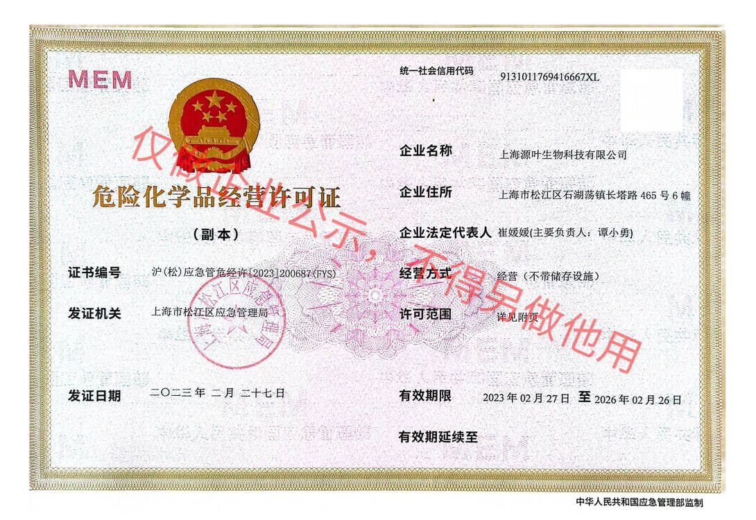 危险品化学品经营许可证(不带存储)
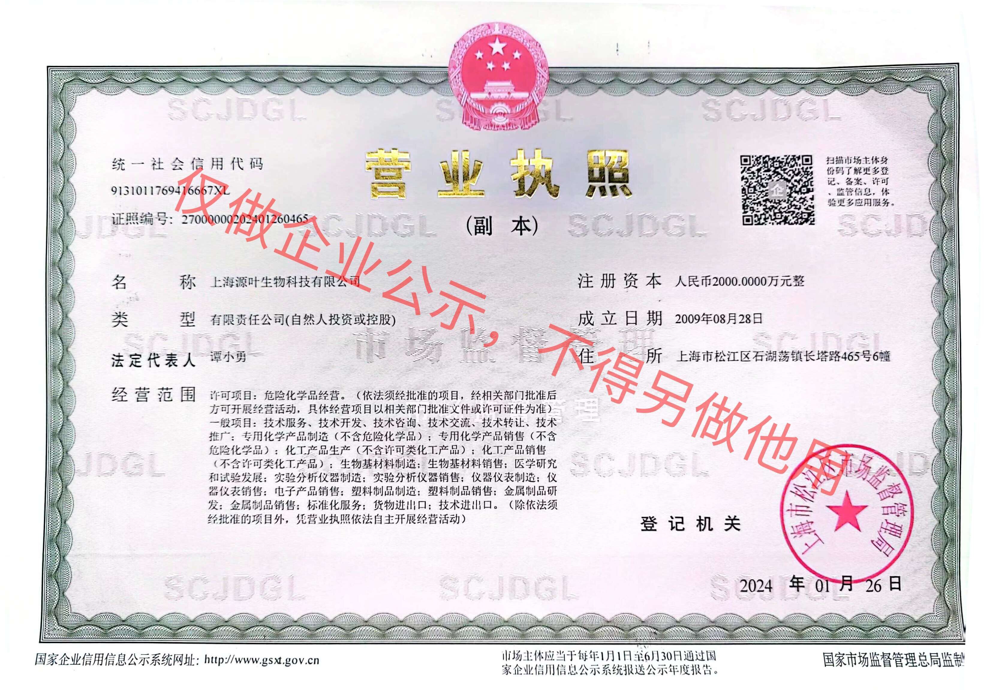 营业执照(三证合一)
